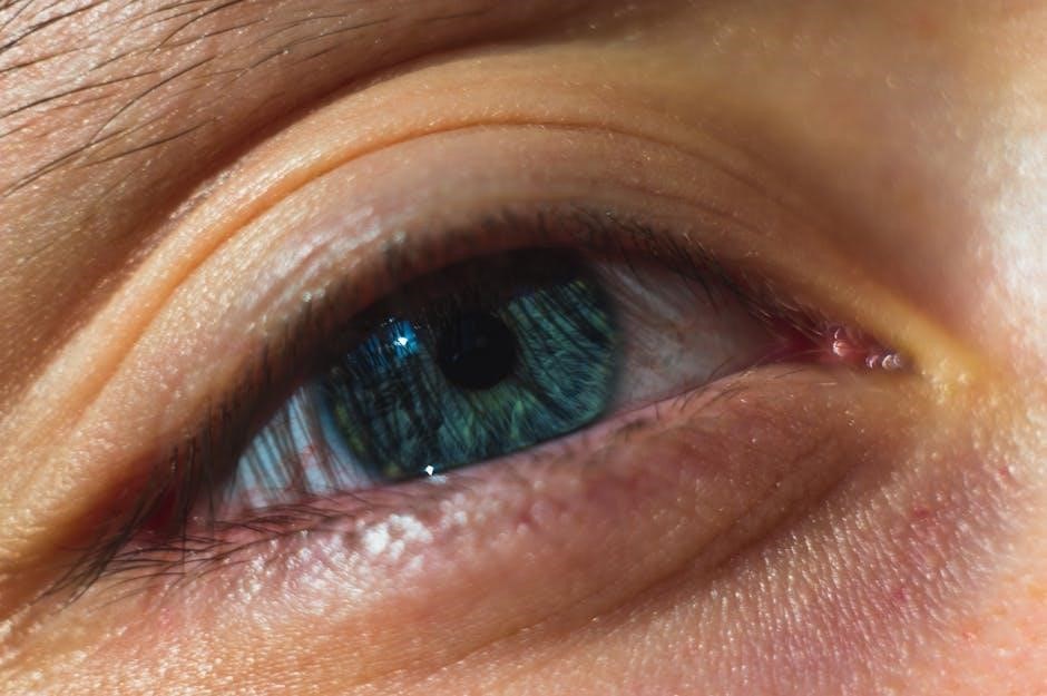
The retina is a light-sensitive tissue crucial for vision. This glossary provides clear definitions of retinal terms, aiding professionals and patients in understanding eye health and conditions effectively.
Overview of the Retina

The retina is a thin, light-sensitive neural tissue lining the back of the eye. It converts light into electrical signals, transmitted to the brain via the optic nerve, enabling vision. Comprising photoreceptor cells (rods and cones), the macula, and ganglion cells, it captures and processes visual details. The macula, at the retina’s center, handles central vision and fine detail. Blood supply comes from the choroid and retinal vasculature, ensuring oxygen and nutrient delivery. Damage to the retina can lead to vision loss, highlighting its critical role in eye health. Understanding its structure and function is essential for diagnosing and managing retinal conditions effectively.
Purpose of a Retina Glossary
A retina glossary serves as a comprehensive resource for understanding technical terms related to retinal health. It simplifies complex terminology, making it accessible to both medical professionals and patients. By defining terms like photoreceptor cells, macular degeneration, and optical coherence tomography, it facilitates clear communication and education. This tool is especially valuable for individuals diagnosed with retinal conditions, helping them comprehend their diagnoses and treatment options. Additionally, it aids healthcare providers in explaining concepts to patients, ensuring informed decision-making. Regular updates keep the glossary current with advancements in retinal research and treatments, ensuring it remains a reliable and authoritative reference for retinal health information.

Anatomy of the Retina
The retina is a thin, light-sensitive tissue lining the eye’s back. It consists of layers, including photoreceptor cells (rods and cones), the macula for central vision, and the optic nerve connecting to the brain.
Structure of the Retina
The retina consists of multiple layers, each serving specific functions. The innermost layer, the inner limiting membrane, separates the retina from the vitreous body. Above it lies the nerve fiber layer, containing axons of retinal ganglion cells. The ganglion cell layer houses the cell bodies of these neurons. The inner plexiform layer facilitates communication between bipolar and ganglion cells. The inner nuclear layer contains amacrine, bipolar, and Müller cells, which support retinal function. The outer plexiform layer connects photoreceptor cells to bipolar cells. The outer nuclear layer holds the nuclei of rod and cone photoreceptors. The photoreceptor layer captures light, while the retinal pigment epithelium supports photoreceptors and absorbs excess light. Finally, Bruch’s membrane separates the retina from the choroid, aiding in nutrient exchange and waste removal. This complex structure enables the retina to process light and transmit visual signals to the brain.
Photoreceptor Cells (Rods and Cones)
Photoreceptor cells, including rods and cones, are specialized neurons in the retina that convert light into electrical signals. Rods are more numerous and sensitive to low light, enabling vision in dim conditions but lacking color perception. Cone cells, fewer in number, detect color and function best in bright light, contributing to sharp central vision. Both cells have an outer segment containing photopigments that absorb light, triggering a biochemical response. Signals from these cells are processed and transmitted to the brain via the optic nerve. Rods and cones are essential for visual acuity and color vision, with cones concentrated in the macula for detailed central vision.
Macula and Its Function
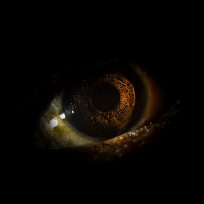
The macula is the central part of the retina responsible for sharp, detailed vision. Located at the back of the eye, it enables activities like reading, driving, and recognizing faces. The macula contains a high concentration of cone cells, which are specialized for color vision and fine detail. Its outer layer is rich in macular pigments like lutein and zeaxanthin, protecting it from harmful blue light. The macula’s function is crucial for central vision, and any damage, such as macular degeneration, can significantly impair visual clarity and independence. Regular eye exams are vital to monitor its health and detect early signs of potential issues.
Optic Nerve and Its Role
The optic nerve is a vital structure connecting the retina to the brain. It transmits visual signals captured by the photoreceptor cells. Damage to the optic nerve can lead to vision loss. The optic nerve is formed by the axons of retinal ganglion cells. These fibers converge at the optic disc and exit the eye. The optic nerve plays a central role in visual processing. Conditions like glaucoma can affect its health. Regular eye exams are essential for early detection of optic nerve issues. Understanding its function is key to maintaining eye health and preventing vision problems. Its role is fundamental in preserving clear sight and overall visual acuity.

Blood Supply to the Retina
The retina receives its blood supply primarily from the central retinal artery and the choroidal circulation. The central retinal artery supplies the inner layers of the retina, while the choroidal circulation, derived from the ophthalmic artery, nourishes the outer layers, including the photoreceptor cells. This dual blood supply ensures the retina’s high metabolic demands are met. Conditions like diabetic retinopathy and macular edema can disrupt this supply, leading to vision problems. Maintaining healthy blood flow is crucial for preserving retinal function and overall vision. Understanding the blood supply to the retina is essential for diagnosing and managing retinal diseases effectively. Proper circulation is vital for eye health.
Common Retinal Conditions
Common retinal conditions include diabetic retinopathy, macular degeneration, retinal detachment, macular edema, and retinal tears. These affect vision and require prompt medical attention for effective management and treatment.
Diabetic Retinopathy
Diabetic retinopathy is a common complication of diabetes, damaging the blood vessels in the retina. It can cause swelling, hemorrhages, and vision loss if left untreated. Early stages, like non-proliferative diabetic retinopathy (NPDR), involve mild damage, while advanced stages, such as proliferative diabetic retinopathy (PDR), lead to abnormal blood vessel growth. Symptoms include blurred vision, floaters, and blind spots. Managing blood sugar levels is crucial for prevention. Treatment options include laser therapy, anti-VEGF injections, and vitrectomy surgery in severe cases. Regular eye exams are essential for early detection and preventing progression to blindness. Prompt medical intervention can significantly improve outcomes for patients with diabetic retinopathy.
Age-Related Macular Degeneration (AMD)
AMD is a progressive eye condition affecting the macula, the central part of the retina responsible for sharp, detailed vision. It is a leading cause of vision loss among older adults. AMD has two forms: dry (atrophic) and wet (neovascular). Dry AMD involves thinning of the macula, while wet AMD is characterized by abnormal blood vessel growth under the retina, leading to fluid leakage and rapid vision loss. Symptoms include distorted or blurred vision and difficulty recognizing faces. Early detection through regular eye exams is crucial. While there is no cure, treatments like anti-VEGF injections and lifestyle changes can manage the condition and slow progression.
Retinal Detachment
Retinal detachment is a serious eye condition where the retina separates from the back of the eye, leading to vision loss. It occurs when fluid seeps under the retina, causing it to pull away. Symptoms include sudden flashes of light, an increase in floating spots, and a curtain or shadow descending over the visual field. If untreated, it can result in permanent vision loss. Treatment typically involves surgery, such as vitreoretinal surgery, or laser therapy to reattach the retina. Prompt medical attention is critical to preserve vision and prevent further damage. Early diagnosis and treatment significantly improve outcomes for patients with retinal detachment.
Macular Edema
Macular edema is a condition characterized by fluid accumulation in the macula, the central part of the retina responsible for sharp vision. This swelling can distort vision, causing blurry or wavy central vision, difficulty reading, and challenges with fine details. It often results from diabetic retinopathy, inflammation, or following eye surgery. Diagnosis typically involves imaging techniques like OCT. Treatment may include anti-VEGF injections, corticosteroids, or laser therapy. Managing underlying conditions like diabetes is crucial to prevent progression. Early intervention can significantly improve visual outcomes, emphasizing the importance of regular eye exams for those at risk of developing macular edema.
Retinal Tears
Retinal tears occur when the retina’s thin tissue develops small breaks or holes, often due to aging or eye injury. If left untreated, tears can lead to retinal detachment, a vision-threatening condition. Symptoms include sudden flashes of light, floaters, or a curtain-like shadow over the visual field. Early detection is critical, as laser or cryotherapy can seal the tears. Regular eye exams are essential for high-risk individuals. Prompt treatment prevents complications, ensuring retinal health and preserving vision. Managing conditions like myopia or diabetes reduces tear risks, highlighting the importance of proactive eye care.

Diagnostic Tools for Retinal Health
Advanced imaging and testing tools like OCT, fluorescein angiography, and fundus photography help detect retinal issues early, ensuring timely treatment and preserving vision. These technologies provide detailed insights into retinal health, enabling accurate diagnoses and effective management of conditions. Regular screenings are essential for maintaining eye wellness and addressing potential problems before they progress. Modern diagnostic tools enhance precision and improve patient outcomes, making them indispensable in retinal care.
Optical Coherence Tomography (OCT)
Optical Coherence Tomography (OCT) is a non-invasive imaging test that uses low-coherence interferometry to capture high-resolution images of the retina. It is widely used to diagnose and monitor retinal conditions such as age-related macular degeneration (AMD), diabetic retinopathy, and macular edema. OCT provides detailed cross-sectional views of the retinal layers, allowing clinicians to measure retinal thickness, detect fluid accumulation, and identify structural abnormalities. This tool is invaluable for early detection and precise monitoring of retinal changes, guiding treatment decisions and improving patient outcomes. Regular OCT scans are essential for managing retinal health and ensuring timely interventions.
Fluorescein Angiography
Fluorescein angiography is a diagnostic imaging technique used to visualize the blood vessels in the retina. It involves injecting a fluorescent dye into a vein, which travels to the retinal blood vessels. A specialized camera captures images as the dye highlights the blood vessels, revealing details about their structure and function. This test is particularly useful for detecting conditions such as diabetic retinopathy, macular degeneration, and retinal vein occlusions. It helps identify areas of leakage, blockage, or abnormal vessel growth, aiding in the diagnosis and management of retinal diseases. Regular use of fluorescein angiography ensures accurate monitoring of retinal health and effective treatment planning.
Fundus Photography
Fundus photography captures detailed images of the retina, documenting its structure for diagnostic and monitoring purposes. Using a specialized camera, this non-invasive technique produces high-resolution images of the retina, including the macula, optic disc, and blood vessels. It is essential for detecting and tracking retinal conditions like diabetic retinopathy, macular degeneration, and glaucoma. Regular fundus photography allows eye care professionals to observe changes over time, enabling early detection of potential issues. This tool is vital for maintaining retinal health and ensuring timely interventions, making it a cornerstone in modern ophthalmology and retina care.

Ultrasound of the Eye
Ultrasound of the eye is a non-invasive diagnostic tool used to examine the retina and other ocular structures when visibility is obscured, such as in cataracts or vitreous hemorrhage. High-frequency sound waves generate detailed images of the eye’s internal structures, aiding in the detection of conditions like retinal detachment, tumors, or optic nerve abnormalities. This technique is particularly useful in emergency situations or when standard imaging methods are inconclusive. It provides valuable insights into retinal health without requiring dilation or direct contact, making it a safe and effective diagnostic option for patients with limited visibility or complex eye conditions.
Treatment and Management
Treatment and management of retinal conditions focus on preserving vision through early detection, lifestyle modifications, and advanced therapies, including medications and surgical interventions tailored to specific diagnoses.
Laser Therapy for Retinal Conditions
Laser therapy is a non-invasive treatment for various retinal conditions. It uses high-intensity light to target and repair damaged areas, such as blood vessels or scar tissue. Common applications include diabetic retinopathy, macular edema, and retinal tears. The laser seals leaking blood vessels, reduces inflammation, and prevents further vision loss. This procedure is precise, minimizing damage to surrounding tissue. Recovery is typically quick, with minimal side effects. Laser therapy is often combined with other treatments, like anti-VEGF injections, for optimal results. It is a critical tool in preserving vision and managing progressive retinal diseases effectively. Early intervention is key for best outcomes.
Anti-VEGF Injections
Anti-VEGF (anti-vascular endothelial growth factor) injections are a cornerstone treatment for retinal conditions like age-related macular degeneration (AMD) and diabetic retinopathy. These drugs block the VEGF protein, which promotes abnormal blood vessel growth and leakage. By halting this process, anti-VEGF therapy prevents vision loss and reduces retinal swelling. Administered directly into the vitreous gel of the eye, these injections are typically repeated monthly or as needed, depending on disease progression. While generally safe, potential side effects include eye discomfort or increased infection risk. Anti-VEGF therapy has revolutionized retinal care, offering a targeted approach to preserve vision and improve outcomes for patients with severe retinal diseases.
Vitrectomy Surgery
Vitrectomy surgery involves the removal of the vitreous gel from the eye, typically performed to repair retinal detachments, clear blood from the vitreous, or treat severe retinal damage. This procedure allows surgeons to access the retina directly, enabling repair of tears, removal of scar tissue, or reattachment of the retina. Modern vitrectomy techniques use small, minimally invasive instruments, reducing recovery time and complications. While the surgery is generally effective, risks include cataract formation, infection, or high pressure within the eye. Recovery requires positioning the head in specific ways to help the retina heal. Vitrectomy is a critical intervention for restoring vision in cases of advanced retinal disease or injury.
Lifestyle Management for Retinal Health

Lifestyle management plays a vital role in maintaining retinal health and preventing vision loss. A balanced diet rich in antioxidants, such as leafy greens and omega-3 fatty acids, supports retinal function. Regular exercise helps control conditions like diabetes and hypertension, which can affect the retina. Avoiding smoking and limiting alcohol consumption reduces the risk of retinal damage. Protecting eyes from UV exposure with sunglasses and managing screen time can also preserve retinal health. Regular eye exams are crucial for early detection of retinal issues. Adopting these habits fosters a healthy retina and reduces the likelihood of conditions like macular degeneration and diabetic retinopathy.

Glossary of Retina-Related Terms
This section provides a comprehensive list of terms related to the retina, offering clear definitions to enhance understanding of retinal anatomy, conditions, and treatments.

A-F
Accommodation: The eye’s ability to focus on objects at varying distances by adjusting the lens’s shape. Diopter: A unit measuring the lens’s refractive power. Macula: The retina’s central part, responsible for sharp, detailed vision. Optic Nerve: Transmits visual signals from the retina to the brain. Retina: The light-sensitive tissue at the eye’s back, essential for vision. Vitreous: The clear gel filling the eye, supporting the retina’s structure. These terms are fundamental to understanding retinal anatomy and function, aiding in diagnosing and treating eye conditions effectively.
G-L
Glaucoma: A condition causing optic nerve damage, often due to high eye pressure. Hyperopia: Farsightedness, difficulty seeing near objects. Iris: The colored part regulating light entry. Laser Therapy: Treats retinal issues by sealing blood vessels or repairing tears. Macular Degeneration: Vision loss from macula deterioration. Myopia: Nearsightedness, difficulty seeing distant objects. Nerve Fiber Layer: Carries visual signals to the optic nerve. These terms are essential for understanding retinal health and conditions, providing clarity for both professionals and patients in eye care diagnostics and treatments.
